
Figure 4. Membrane lipids coordinate signaling in T cells.
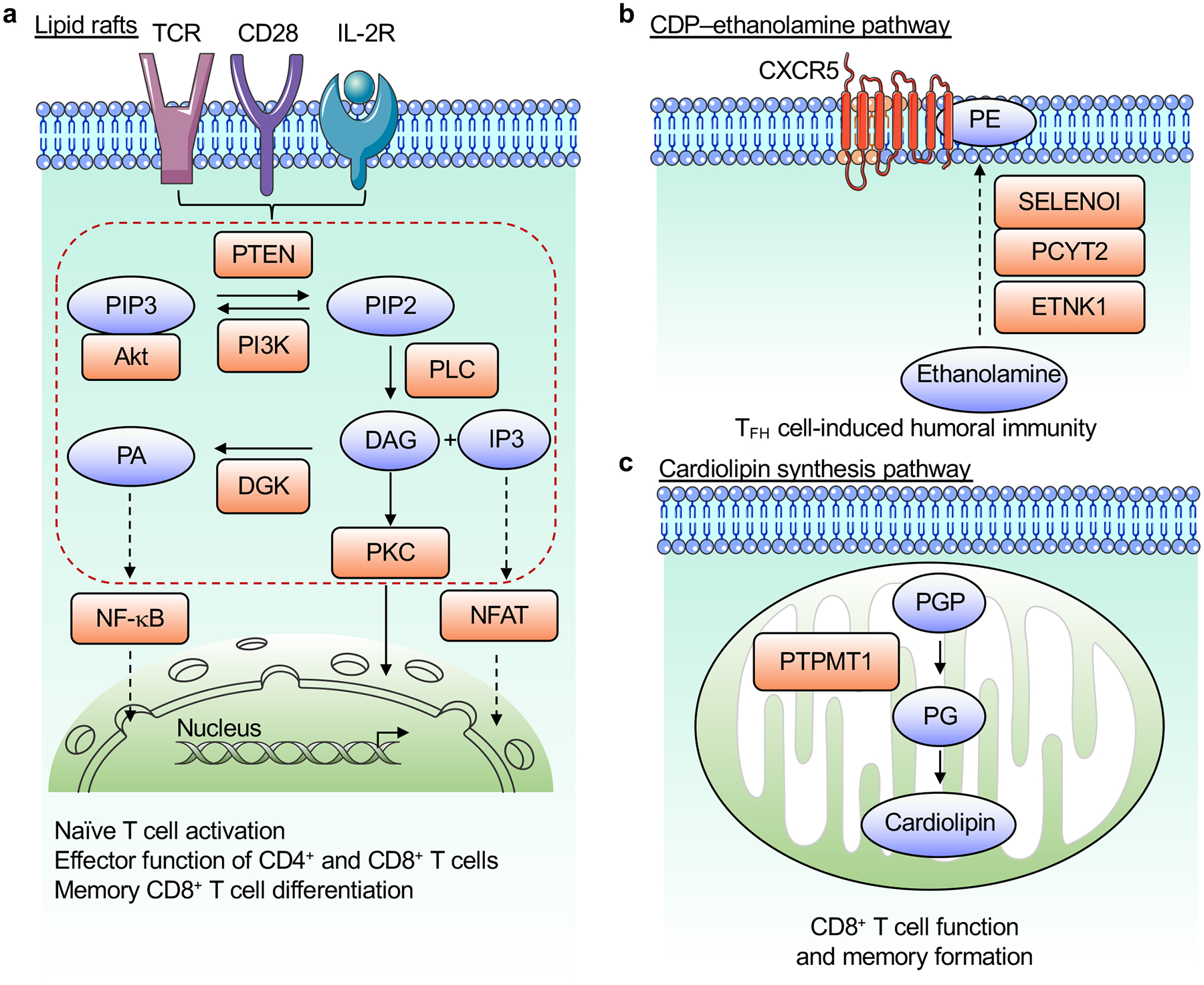
a, Antigens, costimulatory signals and IL-2 stimulation induce PI3K activation that generates PIP3 from PIP2, which is opposed by PTEN. Immunological signals induce PLC activation to produce DAG and IP3 from PIP2. These lipid molecules activate several signaling cascades, including those downstream of PKC, to promote activation of NFAT and NF-κB in the nucleus. DGK opposes DAG-dependent signaling by converting DAG to PA. These lipid-coordinated signaling events play multiple roles in T cell biology. b, De novo PE synthesis via the CDP–ethanolamine pathway, mediated by the enzymes ETNK1, PCYT2, and SELENOI, selectively regulates PE localization to the outer layer of the TFH cell membrane and promotes humoral immunity. c, De novo cardiolipin synthesis in the mitochondria, which depends upon PTPMT1, promotes the function and memory differentiation of CD8+ T cells. DAG, diacylglycerol; IP3, inositol trisphosphate; DGK, diacylglycerol kinase; PA, phosphatidic acid; PIP2, phospholipid phosphatidylinositol 4,5-bisphosphate; PIP3, phosphatidylinositol 3,4,5-triphosphate; PKC, protein kinase C; PLC, phospholipase C; PE, phosphatidylethanolamine; PGP, phosphatidylglycerophosphate; PG, phosphatidylglycerol; PTPMT1, protein tyrosine phosphatase mitochondrial 1.