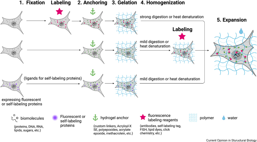
Figure 1.

Three common workflows for expansion microscopy. The first is the original expansion pathway, where samples are fluorescently labeled first and enlarged through anchoring, gelation, homogenization, and expansion steps (top). Alternatively, target biomolecules can be fluorescently labeled after anchoring, gelation, and homogenization (middle). For samples expressing fluorescent proteins, special anchoring crosslinkers and mild homogenization methods can be used to retain the fluorescence signal for imaging after expansion (bottom). Finally, for samples expressing self-labeling protein tags, labeling with ligands that recognize the protein tags is required before anchoring (bottom).
