
Figure 5.
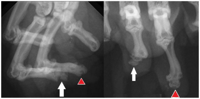
Radiographs of tissue submitted for histopathology. The red arrowhead denotes projections on the bone–cartilage junction comprising bone surrounded by fibrovascular tissue. White arrows denote distal phalanx fragments comprising bone surrounded by fibrovascular tissue