Fig. 3.
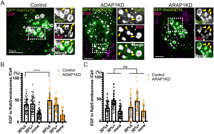
EGF localization in Rab5 endosomes is not altered in ADAP1 and ARAP1KD cells. (A) HeLa cells were transfected with siRNAs as indicated. EGF-Alexa 555 was internalized in HeLa cells for 5 min, washed, and incubated for 40 min under leupeptin. The insets were enlarged, and Rab5 endosomes are indicated with arrowheads. Note that EGF in Rab5 endosomes were dotty. (B) The experiment of control and ADAP1KD cells was repeated three times. More than 15 cells were counted per experiment and more than 45 cells were classified as indicated. The percentages of Rab5-endosome per cell are shown with s.d. Control, n=46; ADAP1KD, n=53. Significance was tested using Mixed-effects analysis. ns, not significant. (C) The experiment of control and ARAP1KD cells was repeated three times. More than 15 cells were counted per experiment and more than 45 cells were classified as indicated. The percentages of Rab5 endosome per cell are shown with s.d. Control, n=60: ARAP1KD, n=44. Significance was tested using the Mixed-effects analysis. ns, not significant, ****P<0.0001. Scale bars: 10 µm.

