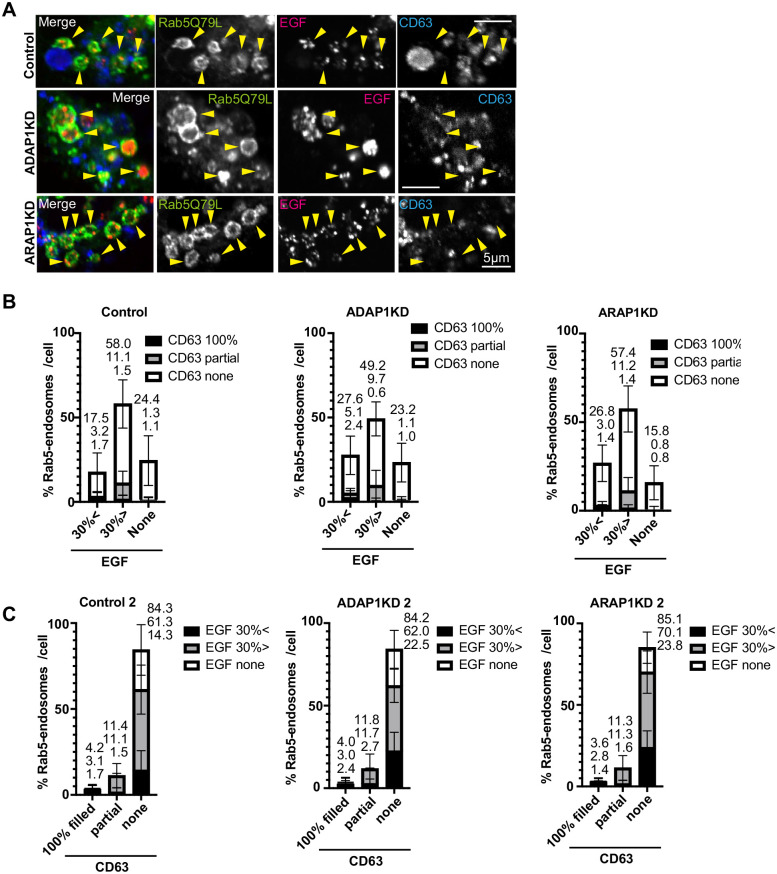
Fig. 5.
EGF and CD63 localization in Rab5 endosomes in HeLa cells. (A) HeLa cells were transfected with each siRNA as indicated, then transfected with GFP-Rab5Q79L (green), and internalized EGF-Alexa 555 for 40 min (red), fixed and stained with anti-CD63 antibody (blue). The representative images of perinuclear region with enlarged endosomes are shown. Rab5 endosomes are indicated with arrowheads. Note that Rab5-endosomes contained dotty signal of EGF, but not CD63. (B) The experiment in A was repeated three times. More than 10 cells were counted per experiment and more than 30 cells were classified as indicated. The percentages of each cell were shown with s.d. The culminated means of each fraction were shown on top of each bar. Control, n=32; ADAP1KD, n=32; ARAP1KD, n=34. (C) The same data set in B were used for the graph of transposed x axis and classification as indicated. Scale bars: 5 µm.

