
Figure 2.
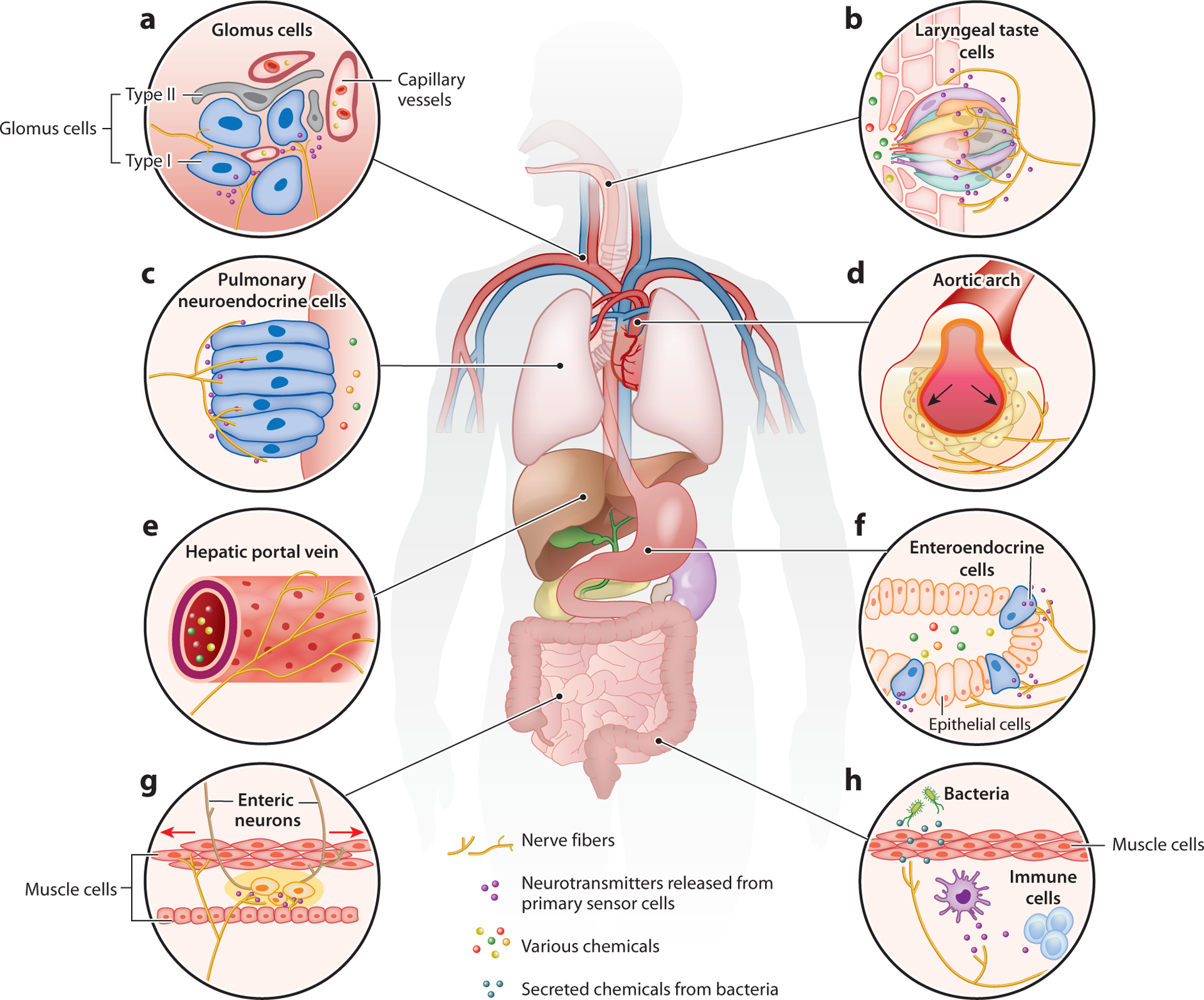
Primary interoceptive sensors. Primary interoceptive sensors can be specialized cells in visceral organs, sensory terminals of peripheral nerves, or sensory neurons in the brain. Depending on the particularity of different types of tissue, specialized organ-resident cells have well-adapted morphology and distribution and express distinct receptors that are specific to the type of stimulus they detect. When activated, these cells release neurotransmitters or neuropeptides to signal nearby peripheral sensory fibers via synaptic or paracrine mechanisms. Alternatively, they can release signaling molecules directly into the bloodstream, allowing for long-range communication throughout the body. Apart from these specialized sensory cells, sensory neurons in the nodose ganglia and dorsal root ganglia also send dense projections and form specialized endings in visceral organs. Examples of primary interoceptive sensor cells include but are not limited to the following. (a) Glomus cells in the carotid body are sensitive to the change in blood oxygen level, carbon dioxide level, and some circulatory hormones. Upon activation, they secrete ATP and a variety of neuropeptides that stimulate afferent sensory fibers. (b) Laryngeal taste cells detect particles and chemicals that enter the airway and secrete neurotransmitters including ATP to nearby vagal fibers. (c) Pulmonary neuroendocrine cells (PNECs) distributed at airway bifurcations respond to mechanical stimuli, hypoxic conditions, and allergens. PNECs also release abundant neurotransmitters and neuropeptides that excite lung-innervating nerves. (d) The aortic arch is densely innervated by the vagus nerve, which detects the change in aortic blood pressure. Arrows indicate that the mechanical force applies to the wall of the aortic arch when blood is pumped from the heart. (e) Sensory nerves on the hepatic portal vein are responsive to multiple hormones related to nutrient absorption. (f) Enteroendocrine cells (EECs) distributed in the epithelium of the gastrointestinal tract sense various contents from ingested food as well as tissue deformation. EECs contain different subtypes and release different signals in response to different stimuli, which modulate activities in the nearby nerves. (g) Sensory enteric neurons are local cells in the gastrointestinal tract that have the capacity to sense gastric volume change and nutrient composition. Enteric neurons can talk to the intraganglionic laminar endings from the vagus nerve and spinal nerve. Intramuscular arrays in the external muscular layers are mechanosensitive vagal endings that fire during gut distension. Arrows indicate stretch of the intestine wall. (h) Tissue barriers and specialized immune cells detect pathogens invading the body. Signal molecules involved in immune responses can also act on periphery sensory nerves. Peripheral sensory fibers can directly be activated by secretions of bacteria within the body.