
Figure 5.
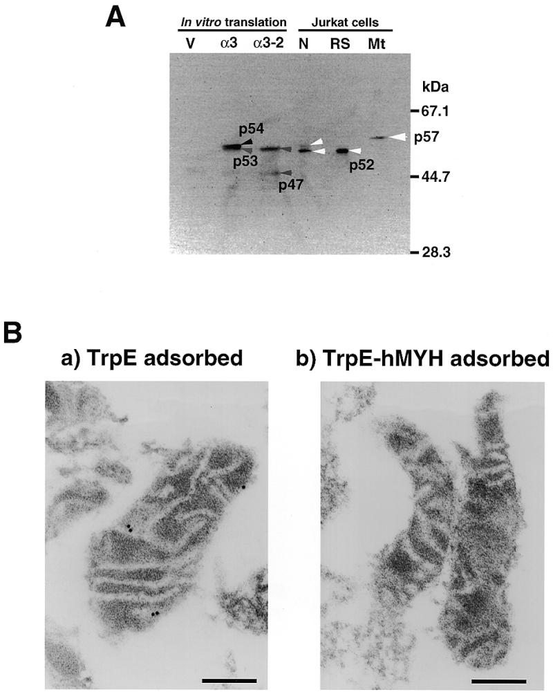
5. Immunological detection of authentic hMYH in human cells. (A) Western blotting. In vitro translation products of the pT7Blue vector (V), pT7Blue:hMYHα3 (α3) and pT7Blue:hMYHα3-2 (α3-2), and isolated nuclei (N) and mitochondria (Mt) (equivalent to 50 µg of protein) from Jurkat cells and partially purified hMYH in the RESOURCE S fraction (RS) were subjected to western blot analysis, using anti-hMYH. (B) Submitochondrial localization of hMYH, determined by electron microscopic immunocytochemistry. After mitochondria had been isolated from Jurkat cells, thin sections (~0.1 µm) were prepared for electron microscopic immunocytochemistry with anti-hMYH preadsorbed with TrpE–Sepharose or with TrpE-hMYH–Sepharose in combination with protein A–gold. Bars indicate 0.2 µm.