
Figure 7.
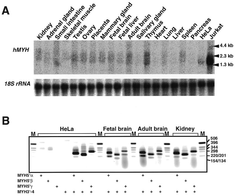
Expression of hMYH mRNAs in various human tissues and cell lines. (A) Northern blot analysis. Total RNAs (16 µg each) extracted from various human tissues and Jurkat and HeLa S3 cells were electrophoresed, transferred to nitrocellulose membrane and probed with 32P-labeled hMYH cDNA. (B) RT–PCR analysis. cDNAs were synthesized from total RNA prepared from HeLa S3 cells, fetal brain, adult brain and kidney, using oligo(dT)18 primer. The cDNAs were amplified using a common 3′ primer (MYH3′-4) and a 5′ primer specific for each type of hMYH mRNA (types 5′α, 5′β and 5′γ). PCR products were analyzed by 1.5% agarose gel electrophoresis. The presence of primers in the reaction mixture is shown by +.