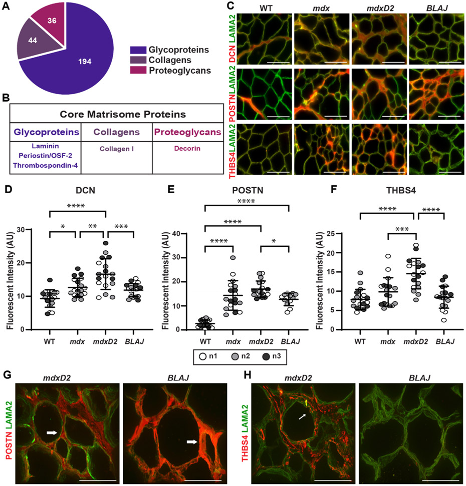
Fig. 4.
Acellular myoscaffolds differentially retained core matrisome proteins decorin, periostin and thrombospondin 4 in different muscular dystrophy models. (A) Core matrisome protein distribution as defined by [60]. (B) Proteins selected for analysis in dECMs. (C) Representative IFM images from WT and mdx, mdxD2, and BLAJ dECMs showing co-staining with antibodies against decorin (DCN), periostin (POSTN), or thrombospondin-4 (THBS4) (red) with laminin-2 (α–2 chain) (LAMA2, green). DCN was observed diffusely throughout dystrophic dECMs from the DBA/2 J background (mdxD2). POSTN was increased in all muscular dystrophy subtypes. THBS4 protein expression, like DCN, was more focally upregulated in specific regions of the dystrophic myoscaffolds and was especially increased in muscular dystrophy models in the DBA/2 J background. Scale bar, 100μm. Quantitation of IFM signal demonstrated a significant increase in mean fluorescent intensity in myoscaffolds (D) DCN (WT 9.7, mdx 13.2, mdxD2 17.3, BLAJ 12.3 AU), (E) POSTN (WT 2.6, mdx 14.3, mdxD2 17.0, BLAJ 12.7 AU), and (F) THBS4 (WT 7.8, mdx 9.8, mdxD2 14.6, BLAJ 8.5 AU). n = 3 independent mice per strain (marked as n1, n2, n3), n = 6 images from 2 scaffolds per mouse. (G,H) 100x Z-stack representative images of POSTN or THBS4 (red) with LAMA2 (green) demonstrate unique protein deposition profiles on the acellular myoscaffolds. POSTN was increased in dysferlin-deficient BLAJ, nearly obliterating the LAMA2 signal (right). In contrast, POSTN signal was interspersed with LAMA2 signal in mdxD2 dECMs (left). In mdxD2 dECM, THBS4 was observed in the matrix in a vesicle-like pattern (thin white arrow). Scale bar, 50μm. Graphs show the mean with SEM bars. * p < 0.05, ** p < 0.01, *** p < 0.005, and **** p < 0.001 by one-way ANOVA.

