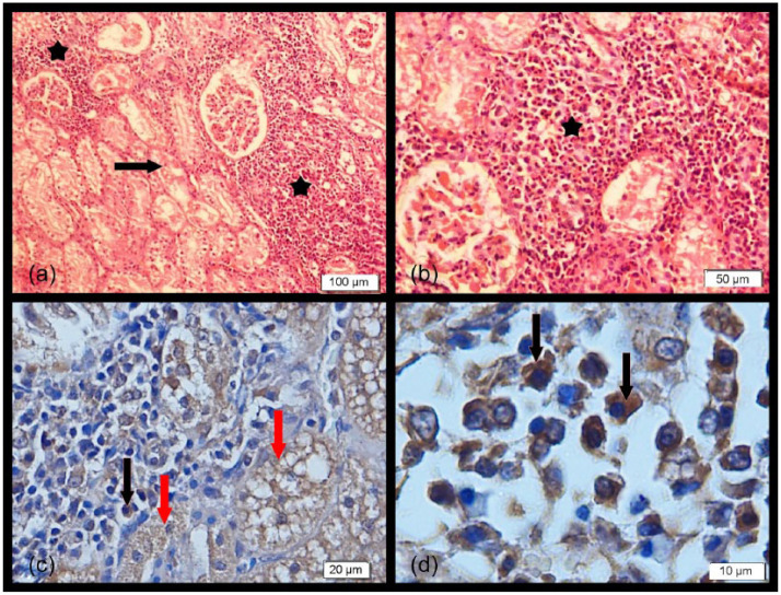
Figure 1.
(a,b) Prominent mononuclear cell infiltrates composed of lymphocytes and plasma cells (stars) and severe granular and vacuolar degeneration in the epithelial cells of tubules (arrow) (haematoxylin and eosin). (c,d) Kidney sections showing intensely positive intracytoplasmic staining of tubular epithelial cells (red arrows) and mononuclear cells (black arrows) (immunohistochemistry)

