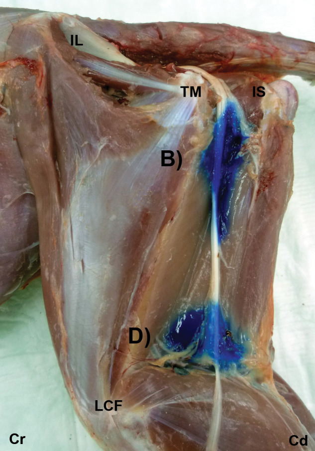
Figure 3.

Gross dissection images after US-guided injection of 1 ml ink at the glutea caudal (B) and poplitea (D) approaches. The ink is observed around the ScN. IL = ala ossis ilii, TM = trochanter major of os femoris, IS = tuber ischiadicum, LCF = condylus lateralis of os femoris, Cr = cranial, Cd = caudal
