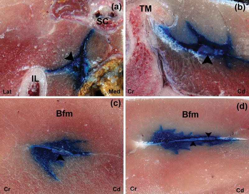
Figure 4.
Cross-sectional images after US-guided injection of 1 ml ink at the glutea cranial (a), glutea caudal (b), femoris (c) and poplitea (d) approaches. The ink is observed around the ScN (arrow head). IL = ala ossis ilii, TM: = trochanter major of os femoris, Bfm = biceps femoris muscle, Sc = os sacrum, Lat = lateral, Med = medial, Cr = cranial, Cd = caudal

