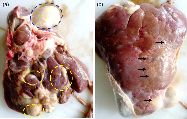
FIGURE 1.

The affected liver of the goat. (a) visceral view. Blue circle, gallbladder; yellow circle, cyst‐like structure in the liver that contained flukes and tarry coloured fetid materials. (b) parietal surface of the liver. Arrows indicate grossly white patches of fibrosis.
