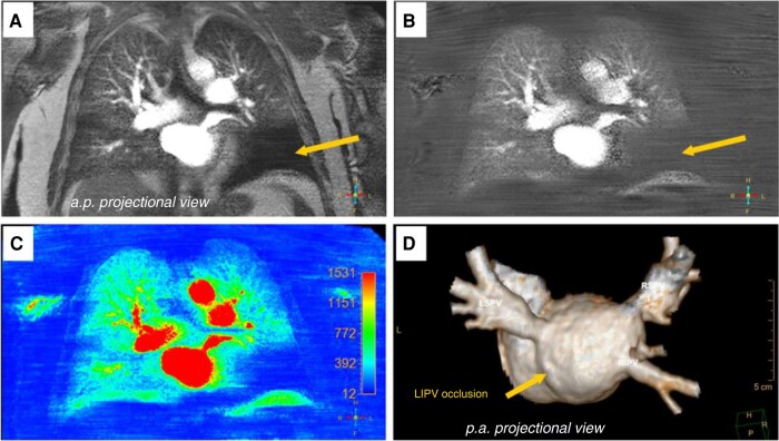
Figure 11.
Pre-interventional CMR (cardiovascular magnetic resonance) pulmonary perfusion imaging and angiography for detection and characterization of pulmonary vein stenosis. A, B, C) CMR pulmonary perfusion imaging (anterior–posterior view) depicted a perfusion deficit of the left lower lung lobe (A still frame of original dynamic pulmonary perfusion; B still frame of dynamic pulmonary perfusion after background stationary tissue subtraction; C corresponding pseudo-coloured parametric map of quantitative CMR pulmonary perfusion analysis with SI maximum enhancement as the quantitative measure. D) Three-dimensional contrast-enhanced CMR angiography (posterior–anterior projectional view) revealed total ostial occlusion of the left lower pulmonary vein (arrow). CMR, cardiac magnetic resonance; SI, signal intensity.

