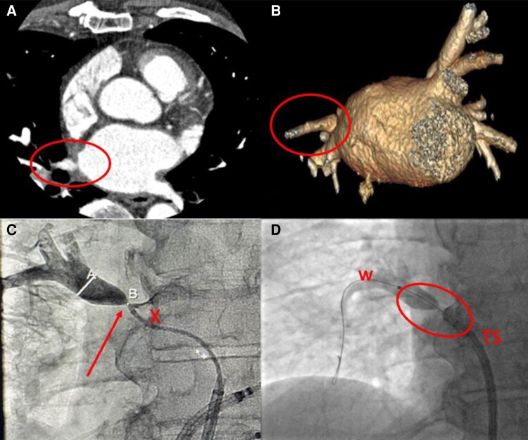
Figure 9.
A patient with history of a prior PV isolation procedure and history of recurrent pneumonia in the right superior lobe following the procedure. A) CT showing high-degree stenosis of the right superior PV (circle), B) 3D reconstruction of the left atrium demonstrating a high-degree ostial stenosis of the right superior PV (circle), C) angiography of the right superior PV after transseptal access through a coronary diagnostic catheter (x) showing high-degree stenosis (arrow) (A indicates diameter of PV after stenosis, B site of stenosis), D) contrast injection into right superior PV after wiring (w) of stenosis (red circle) with transseptal sheath close to PV ostium. CT, computed tomography; PV, pulmonary vein.

