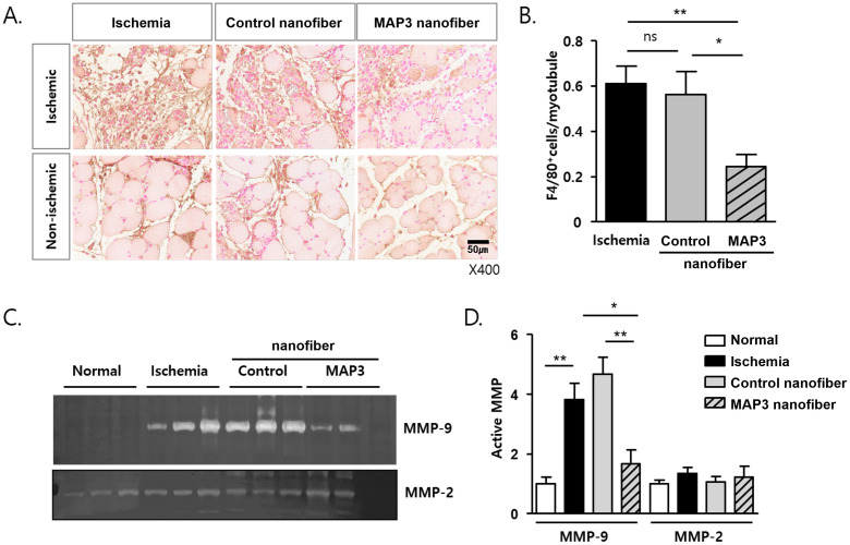
Fig 3. MAP3 ameliorates macrophage infiltration and MMP9 activity.
(A) Representative images of immunohistochemical stain with F4/80 and eosin from paraffin-sections of day 4 post-ischemia. Both of ischemic wound area and non-ischemic area were observed. (B) F4/80 positive macrophages of sections from non-ischemic area were counted and presented as numbers of F4/80+/myotubules. (C) Gelatin-zymography were performed with same amount of proteins from tissue homogenates of day 4 post-ischemia. Representative MMP-9 and MMP-2 gelatinolytic images were shown. Age matched normal mice tissues were used (Normal). (D) MMP-9 and MMP-2 activities were analyzed with the clear band and the relative activities to normal were shown as mean ± standard error of the mean. *p<0.05, **p<0.01. MAP3: 3-methylaminopropyltrimethoxysilane; MMP: Matrix metalloproteinase.

