Figure 5.

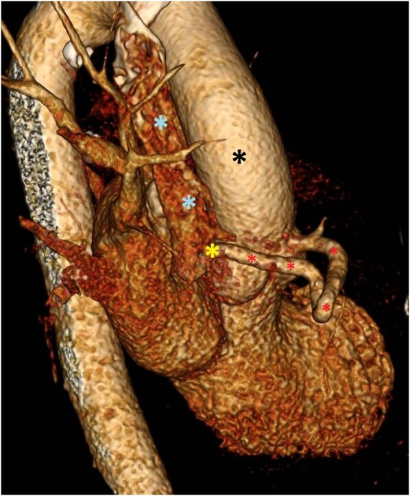
EKG-gated CTA chest, side view. This image depicts a 3D reconstruction from EKG-gated CTA. The aorta (black asterisk) and SVC (blue asterisk) are visible, with an abnormal RCA–SVC connection (yellow asterisk) via a fistula (red asterisk).

EKG-gated CTA chest, side view. This image depicts a 3D reconstruction from EKG-gated CTA. The aorta (black asterisk) and SVC (blue asterisk) are visible, with an abnormal RCA–SVC connection (yellow asterisk) via a fistula (red asterisk).