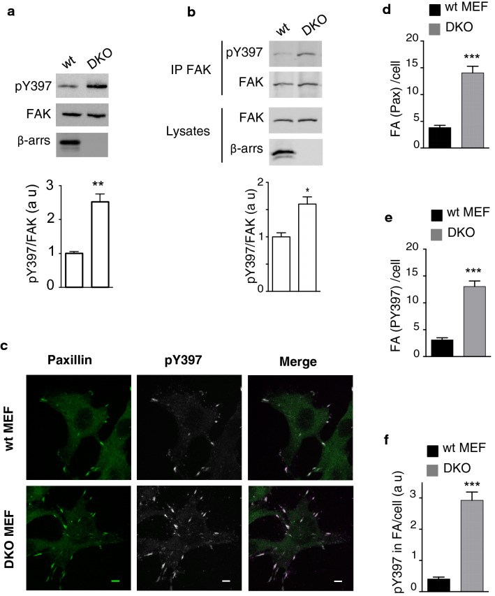
Fig. 1.
β-arrs regulate FAK autophosphorylation and focal adhesion number. a Adherent wt and β-arr1−/−/β-arr2−/− (DKO) MEF cells were serum starved, lysed, and analysed for FAK-pY397, FAK, and β-arrs by immunoblotting. The mean ± s.e.m. of pY397/FAK values, calculated from three independent experiments, was normalized to the value obtained in wt MEFs. (**P value < 0.01, t test). b Immunoblotting for FAK-pY397 and total FAK was performed after IP FAK from lysates of serum-deprived MEF cells held in suspension for 60 min at 37 °C. Quantification (lower panel, *P < 0.1, t test) was performed as in a. c Confocal images of serum-starved MEF cells fixed and stained for paxillin (green) and pY397-FAK (grey). Merge is shown as paxillin (green) and pY397-FAK (magenta). Scale bar = 5 µm. FA number per cell indicated by paxillin (d) or pY397-FAK staining (e) and pY397-FAK intensity staining in focal adhesions (FA) per cell (f) were quantified in MEFs from c. For each condition, 35–40 cells from different coverslips were quantified (***P < 0.001, t test)

