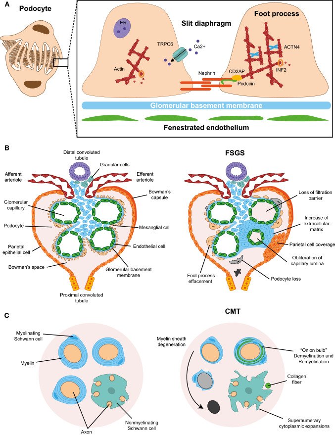
Fig. 2.
Phenotypes caused by pathogenic mutations. a Schematic of podocyte structure (left panel) and magnification of the slit diaphragm formed between adjacent pedicels, indicating some of the proteins whose mutation causes FSGS (right panel). b Structure of a glomerulus in control (left panel) and in patients with FSGS (right panel), indicating the various glomerular structures and the typical alterations found in FSGS. c Myelinating and nonmyelinating cells in control (left panel) and in patients with FSGS + CMT caused by INF2 mutation (right panel). In the latter, the myelin sheath formed by Schwann cells degenerates progressively, leading to the appearance of “onion bulbs” and unmyelinated axons, and the nonmyelinating Schwann cells change their morphology and display supernumerary extensions

