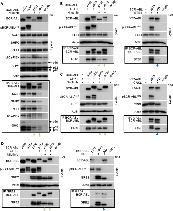
Fig. 3.
The BCR–ABL signaling complex is preserved after nilotinib treatment. 293T cells were transfected with p190 and p210 BCR–ABL alone (a) or together with STS1 (b), CRKL (c) and GRB2 (d). BCR–ABL was immunoprecipitated (IP) and binding of interaction partners was analyzed by western blot. The SHC1 isoforms are indicated (p46, p52, p66). Empty, transfection with empty plasmid. BCR–ABL kinase activity was determined by detecting autophosphorylation (p) at Y412. Note the co-immunoprecipitation of SOS1, SHIP2, cCBL, SHC1, STS1, CRKL and GRB2 with BCR–ABL in cells treated with nilotinib (green arrows). Also note the co-immunoprecipitation of STS1, CRKL, and GRB2 with kinase-dead (KD) BCR–ABL (blue arrows). Data are representative of three independents experiments (n = 3). Actin serves as a loading control in cell lysates used for IP

