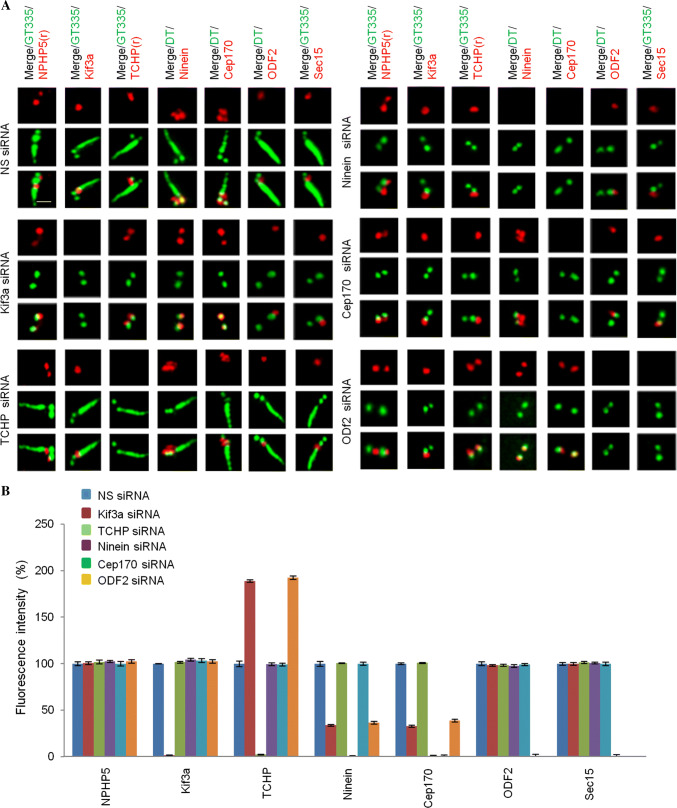
Fig. 6.
Hierarchical assembly of BF. a Quiescent RPE-1 cells transfected with NS (non-specific) or the indicated siRNAs targeting SDA/BF components (Kif3a, TCHP, ninein, Cep170, ODF2) were stained with the indicated antibodies. Scale bar, 1 µm. b Fluorescence intensities of various proteins at the centrosome were quantitated and set to 100% in NS siRNA-transfected cells. For quantitation, at least 20 cells for each condition were analyzed, and the mean and standard error of three independent experiments are presented

