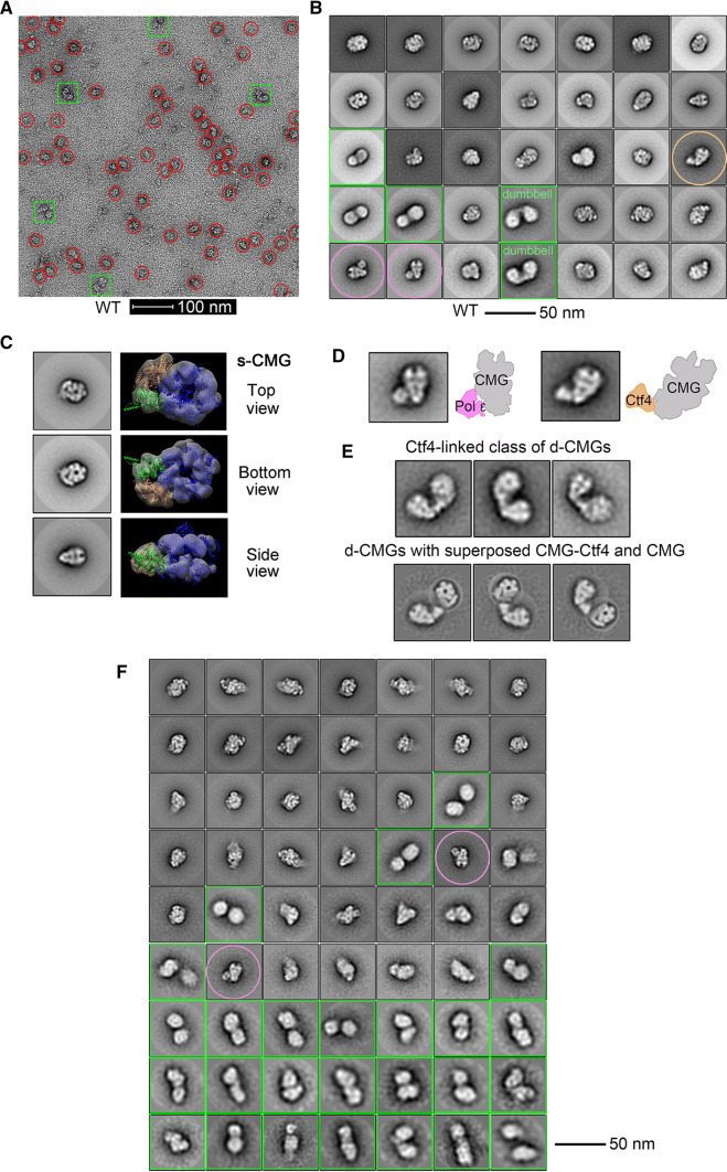
Fig. 6.
Single-particle EM analysis of the negatively stained CMG complexes. a A representative electron micrograph of the endogenous CMG-containing complexes (fractions 17–22) isolated from the S-phase cells of CDC45-5FLAG (LL85, Table S1) through the same purification procedure as described in Fig. 4. The putative single (s-CMG) and double (d-CMG) CMG particles are highlighted by red circles and green squares, respectively. b 2D class averages of all types of CMG-containing particles (38,787 in total). The putative CMG-DNA pol ε and CMG-Ctf4 particles are circled with pink and tawny, respectively. c S-CMG particles with top/bottom and side views. d S-CMG particles containing DNA Pol ε or Ctf4. e The dumbbell-shaped d-CMG particles (824 among total 6445 d-CMGs) with computationally superposed CMG-Ctf4 and CMG. f 2D class averages of the CMG particles (43,820 in total) purified endogenously from the S-phase ctf4Δ cells (LL-163, Table S1). The particles of putative d-CMG and CMG-DNA pol ε are labeled in green squares and pink circles, respectively

