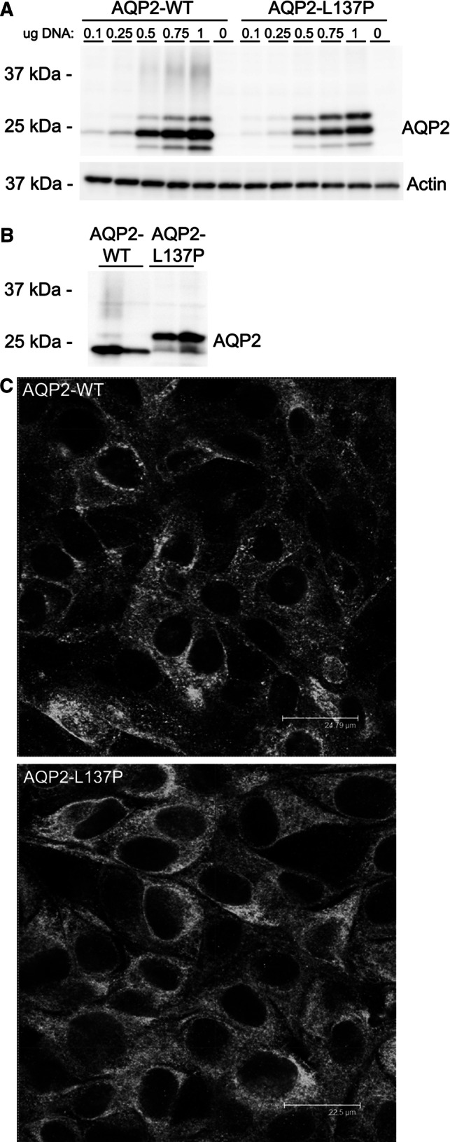
Fig. 1.

Compared to AQP2-WT, the AQP2-L137P mutant has a different molecular mass and subcellular distribution when expressed in kidney epithelial cell lines. a Immunoblotting of AQP2 on cell lysates isolated from HEK293 cells transiently transfected with AQP2-WT and AQP2-L137P constructs. Various amounts of plasmid DNA were used for transfection. AQP2-WT is mainly detected as a ~ 25 kDa band, but AQP2-L137P is distributed evenly between ~ 25 kDa and 27 kDa forms. Actin is used as a loading control. b Immunblotting of cell lysates from MDCK cells stably expressing AQP2-WT or AQP2-L137P revealed a similar tendency for the mutant protein to exist as a higher molecular weight form compared to WT-AQP2. Upon longer exposure a complex glycosylated smear could be observed for the AQP2-WT-expressing cells. Each lane represents a lysate from a different stable cell line. c Confocal images of AQP2 distribution in MDCK cells stably expressing AQP2-WT or AQP2-L137P
