Fig. 2.
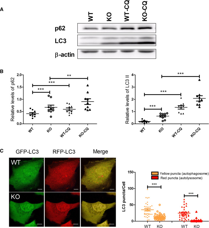
EI24 deficiency impairs the autophagy flux. a U-2 OS cells were treated with or without 100 μM chloroquine (CQ) for 6 h. Total protein was extracted and separated by SDS-PAGE and immunoblotted with anti-p62 and anti-LC3 antibodies. b Quantification analysis of p62 and LC3II, normalized to β-actin. c U-2 OS cells were transfected with the mRFP–GFP–LC3 plasmid, and the left panel represents fluorescent images of U-2 OS cells acquired by confocal microscopy. The right panel shows the quantification of the number of autophagosome (yellow) and autolysosome (red) puncta in WT and EI24 KO U-2 OS cells. WT (n = 34), KO (n = 40). Bar = 10 μm, **p < 0.01, ***p < 0.001

