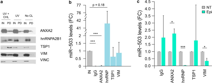
Fig. 3.
Epirubicin disrupts the interaction between miR-503 and VIM and hnRNPA2B1. a HUVECs (30·106 cells) were transfected with miR-503-biotin (10 nM). The following day, the cells were crosslinked with DTSSP and UV (UV + CHL), only UV (UV) or not subjected to any crosslinking (No CL). HUVECs lysates were incubated with streptavidin beads. Both input (IN) (cellular lysate) and pull-down fractions (PD) were separated by SDS-PAGE and revealed by western blotting using indicated proteins. Input (IN) = 1% of cell lysate. Vinculin was used as loading control. b, c HUVECs were transfected with miR-503 (10 nM). 48 h later, immunoprecipitation assays were performed using the indicated antibodies or an IgG control and the levels of miR-503 were evaluated by qPCR. Plots show b fold change of miR-503 in the immunoprecipitated (IP) vs input fractions (IN) in non-treated cells and c fold change of immunoprecipitated miR-503 in epirubicin-treated (EPI) vs non-treated cells (NT). Plots represent mean and SEM from three independent experiments,*p < 0.05; **p < 0.01, ***p < 0.001. IN = 1% of the cellular lysate before immunoprecipitation

