Fig. 3.
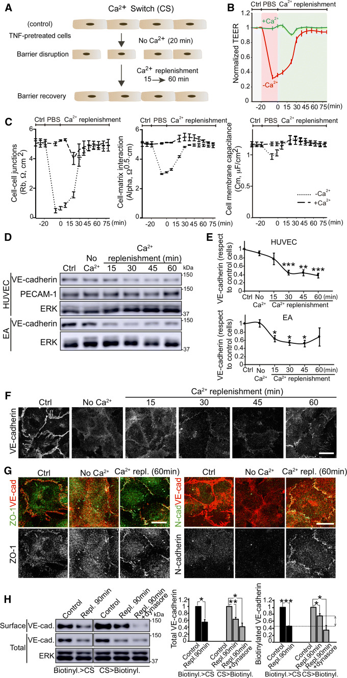
An intracellular VE-cadherin pool partially compensates surface VE-cadherin decrease upon acute barrier disruption a, b HUVECs were cultured at confluence for 48 h on dishes pre-coated with fibronectin, starved for 12 h and then stimulated with 10 ng/ml TNFα for 24 h before performing a Ca2+ switch (CS) assay, which induces a transient and acute disruption of the cell monolayer in absence of Ca2+ and a subsequent recovery upon Ca2+ replenishment with EBM-2 endothelial culture medium. b ECIS analysis of the transient effect of Ca2+ switch on TEER. PBS with no Ca2+ induces a strong decrease in TEER, whereas PBS containing 1 mM Ca2+ has no effect. c ECIS mathematical modelling of the TEER analysis in (b) yields three values: Rb values, which reveal changes in paracellular permeability (left), Alpha values, which are proportional to changes in cell–matrix interactions (center), and Cell membrane capacitance (right) [21]. d Analysis of VE-cadherin expression levels during the CS assay in HUVECs and EA.hy926 (EA) detected by western blot. e Graphs show VE-cadherin expression levels with respect to control cells from at least three independent experiments. *p < 0.05; **p < 0.01; ***p < 0.001. f VE-cadherin distribution in HUVECs during the CS assay. Scale bar, 20 μm. g ZO-1 and N-cadherin distribution with respect to VE-cadherin staining during the CS assay. Scale bar, 20 μm. h TNFα-pretreated cells were biotinylated before or after a CS assay. Control cells and cells recovering after 90 min of Ca2+ replenishment were lysed and biotinylated proteins isolated by pull-down with NeutrAvidin-agarose beads. Graphs show the quantification of total and surface VE-cadherin levels from three independent experiments. *p < 0.05; **p < 0.01; ***p < 0.001. ERK was immunoblotted as a loading control. Double arrowhead shows differences in surface VE-cadherin during Ca2+ replenishment when cells were biotinylated before and after CS, which correspond to the surface localization, after barrier reformation, of VE-cadherin molecules that were not at the cell surface before the CS assay (right graph). To test whether VE-cadherin localization at the cell surface requires intracellular trafficking, parallel experiments of CS > biotinylation where performed in the presence of Dynasore during Ca2+ replenishment. See also Fig. S3

