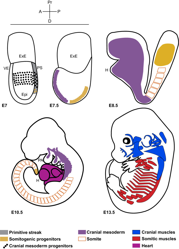
Fig. 1.
Schematic illustrating the origin and derivatives of cranial mesoderm. Cartoons of mouse embryos from mid-gastrula stage showing cranial and somitic mesoderm progenitors and their muscle derivatives. Embryonic day E7 all mesodermal progenitors emerge from the posterior end, where the primitive streak is formed. E7 and E7.5 cranial mesoderm migrates to the anterior pole, while somitogenic progenitors develop on the posterior side. E8.5 and E10.5 lateral plate of cranial mesoderm forms the heart and the paraxial component spreads as a non-segmented mesenchyme and eventually patterns into streams entering the pharyngeal arches. This contrasts with posterior paraxial mesoderm forming somites. E13.5 the pharyngeal mesodermal core contributes to different cranial skeletal muscle groups (in blue). Somite-derived skeletal muscles are shown in red. VE visceral endoderm, ExE extra-embryonic ectoderm, Epi epiblast, PS primitive streak (marked in grey), H heart, PA pharyngeal arches, A anterior, P posterior, Pr proximal, D distal. E7, 7.5 (based on evidence from [40]), E8.5 (based on evidence from Mesp1-cre/R26R; [153]), E10.5 [129] and E13.5 [53]

