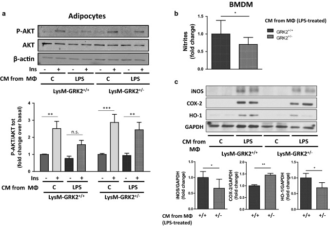
Fig. 7.
Effect of the conditioned medium from thioglycollate-elicited peritoneal macrophages (TEPMs) isolated from either LysM-GRK2+/+ or LysM-GRK2+/− mice on adipocytes and naïve bone-marrow-derived macrophages (BMDM). a 3T3L1 adipocytes were stimulated for 24 h with the conditioned medium (CM) obtained from TEPMs isolated from either LysM-GRK2+/+ or LysM-GRK2+/− mice previously treated or not with 1 µg/ml LPS for 6 h. 24 h later, 3T3L1 adipocytes were stimulated with insulin (Ins,10 nM) for 10 min and cell lysates were subjected to WB using antibodies against total and phosphorylated AKT (Ser473) and β-actin. Representative immunoblots (upper panel) and densitometric analysis (lower panel) of three independent experiments are shown. Results are expressed as fold change over basal (3T3L1 cells stimulated with CM from TEPMs non-stimulated with LPS and in the absence of insulin). Statistical significance was analyzed by two-way ANOVA followed by Bonferroni post-hoc test (**p < 0.01; ***p < 0.005). b Relative nitrite levels produced by BMDM upon 24 h-stimulation with CM obtained from TEPMs isolated from either LysM-GRK2+/+ or LysM-GRK2+/− mice previously treated with 1 µg/ml LPS for 6 h. Results are expressed as fold change of nitrite concentration in the supernatant of BMDM stimulated with the CM of LPS-treated GRK2+/− macrophages over control (supernatant of BMDM stimulated with the CM of LPS-treated GRK2+/+ macrophages). c Representative immunoblots (upper panel) and densitometric analysis normalized to GAPDH levels (lower panel) of iNOS, COX-2, HO-1 and GAPDH from BMDM exposed for 24 h to CM from LysM-GRK2+/+ or LysM-GRK2+/− TEPMs stimulated or not with 1 µg/ml LPS for 6 h.. Graphs represent fold change over control (supernatant of BMDM stimulated with the CM of LPS-treated GRK2+/+ macrophages). Statistical significance was analyzed by unpaired two-tailed t test (*p < 0.05; **p < 0.01)

