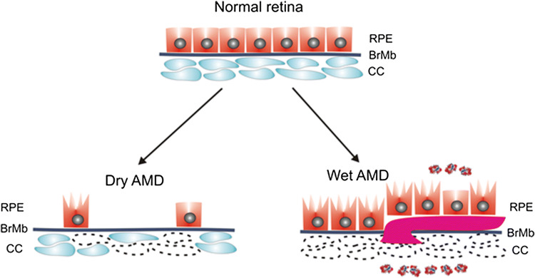
Fig. 1.
Dry and wet AMD. The normal retina contains photoreceptors (not presented here) that are in contact with the retinal pigment epithelium (RPE)/Bruch’s membrane (BrMb)/choriocapillaris (CC) complex, which is underlined by large choroidal blood vessels (not shown). In dry age-related macular degeneration (AMD), RPE cells are progressively lost with disease progression. Some of the RPE cells in dry AMD may be damaged and some CC are lost (broken line). In wet AMD, CC are almost completely lost, become hypoxic and produce hypoxia-inducible growth factors, including vascular endothelial growth factor (VEGF, multi-color small objects) that induce formation of choroidal neovascularization (CNV) presented as a large purple vessel, penetrating BrMb and RPE. In advanced stage of wet AMD, almost all RPE cells are damaged, but they still reside on the top of CNV vessels [5]

