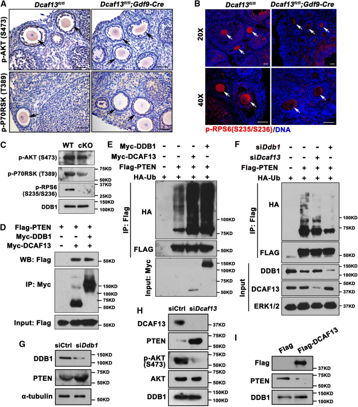
Fig. 6.
CRL4DCAF13 targets PTEN for polyubiquitination and degradation. a–c Immunohistochemistry (a), immunofluorescence (b), and western blot (c) results showing the level of phosphorylated AKT (Ser473), p70 ribosome S6 kinase (P70RSK-Thr389), and RPS6 (Ser245/236) in oocytes of 4-week-old mice with indicated genotypes. Arrows indicate growing oocytes. Scale bars, 50 μm. d Co-immunoprecipitation result showing PTEN interaction with DCAF13 and DDB1. HeLa cells were co-transfected with Flag-PTEN and Myc-DCAF13 or Myc-DDB1 expression plasmids for 24 h. Target proteins were immunoprecipitated using anti-Myc beads and subjected to western blotting with FLAG and Myc antibodies. Input cell lysates were immunoblotted with an anti-Flag antibody to determine the expression of PTEN. e HeLa cells transiently transfected with plasmids encoding the indicated proteins were lysed and subjected to immunoprecipitation with an anti-HA affinity gel. Input cell lysates and precipitates were immunoblotted with antibodies against FLAG, HA, and MYC. f Co-IP followed by western blotting showing PTEN polyubiquitination in control HeLa cells and those transfected with Dcaf13 or Ddb1 siRNAs. g Western blotting results showing the levels of PTEN in oocytes microinjected with control siRNA (siCtrl) or siDdb1.α-Tubulin was blotted as a loading control. h Western blotting results showing the levels of PTEN, AKT, and phosphor-AKT (Ser473) in oocytes after microinjected with siControl or siDcaf13 and further cultured for 36 h. DDB1 was blotted as a loading control. i Western blotting results showing the levels of PTEN in oocytes after microinjected with Flag-vector or Flag-DCAF13 mRNA and further cultured for 24 h. DDB1 was blotted as a loading control. Sizes (kDa) of protein markers are indicated on the right in all figures

