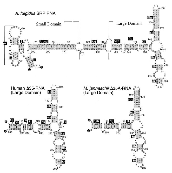
Figure 1.
Secondary structures of A.fulgidus SRP RNA and Δ35 RNAs of H.sapiens and M.jannaschii. Secondary structures are shown with base pairings supported by comparative sequence analysis of SRP RNA sequences in the SRP database (14). 5′- and 3′-ends of RNA molecules are labeled as such; helices are numbered 2–8 according to the nomenclature of Larsen and Zwieb (25). Base paired sections of helices 5, 6 and 8, including regions of coaxial stacking (48), are highlighted in gray and labeled in reverse print with suffices a–k in helix 5, and a–c in helices 6 and 8. Residues are numbered in 10-nucleotide increments and marked with dots in 10-nucleotide increments in reference to full-length molecules.

