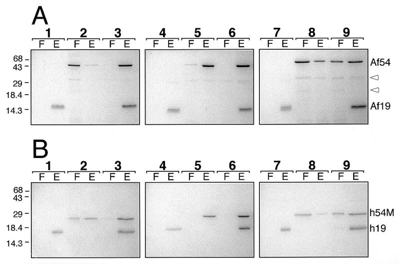
Figure 6.

Assembly with proteins of A.fulgidus or human SRP. (A) Binding of 1 µg Af-SRP19 and 3.8 µg Af-SRP54 polypeptides to 10 µg Af-SRP RNA (lanes 1–3), 10 µg Mj-SRP RNA (lanes 4–6) or 10 µg human SRP RNA (lanes 7–9). (B) Binding of 1.3 µg human SRP19 and 2 µg human SRP54M polypeptides to 10 µg Af-SRP RNA (lanes 1–3), 10 µg Mj-SRP RNA (lanes 4–6) or 10 µg human SRP RNA (lanes 7–9). Each panel shows polypeptides in the flowthrough (F) and eluate (E) as determined in DEAE binding assays (see Materials and Methods) followed by electrophoresis of polypeptides on 15% polyacrylamide gels and staining with Coomassie blue. Lanes 1, 4 and 7, addition of SRP19; lanes 2, 5 and 8, addition of Af-SRP54 or human SRP54M; lanes 3, 6 and 9, addition of both Af-SRP19 and Af-SRP54 (A) or human SRP19 and human SRP54M (B). Mobilities of Af-SRP54 (Af54), Af-SRP19 (Af19), human SRP19 (h19) and human SRP54M (h54M) polypeptides are indicated on the right. Open arrowheads in (A) mark two proteolytic products of Af-SRP54. Migration distances of molecular weight markers with sizes in kDa are indicated on the left.
