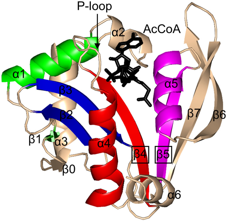
Fig. 5.
Cartoon representation of the structure of H. pylori PseH in complex with AcCoA (PDB ID: 4RI1 [129]). The motifs that are conserved across all GNAT enzymes are coloured as follows: motif C—green, motif D—blue, motif A—red, motif B—magenta. Non-conserved N-terminal and C-terminal regions are coloured wheat. The AcCoA cofactor is drawn in black using a stick representation

