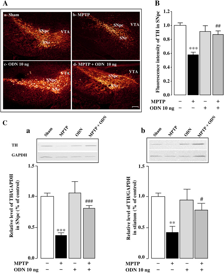
Fig. 2.
ODN reverses the reduction of tyrosine hydroxylase expression in MPTP-treated mice. A Representative images of tyrosine hydroxylase (TH) immunostaining in the substantia nigra pars compacta (SNpc) 7 days after treatment with NaCl (Sham; A-a), MPTP (3 × 20 mg/kg; A-b), ODN (10 ng/10 µl; A-c) and MPTP + ODN (A-d). Scale bar = 100 µm SNpc (substantia nigra pars compacta), SNr (substantia nigra pars reticulate), VTA (ventral tegmental area). B Relative TH immunofluorescence intensity of surviving dopaminergic neurons in the SNpc 7 days after MPTP treatment alone or with ODN. C Densitometric analysis of TH protein levels in the SNpc (C-a) and striatum (C-b) of sham-, MPTP-, ODN-, and MPTP + ODN-treated mice. Digital photographs illustrate the expression of TH after immunoblotting, and graphs display the relative abundance of TH measured by densitometry of the bands obtained in immunoblots and standardized with GAPDH. All values are expressed as mean ± SEM from 5 animals and statistical analysis was conducted by ANOVA followed by Bonferroni’s test. **P < 0.01, ***P < 0.001 vs saline-treated mice; # P < 0.05, ## P < 0.01, ### P < 0.001 vs MPTP-treated mice

