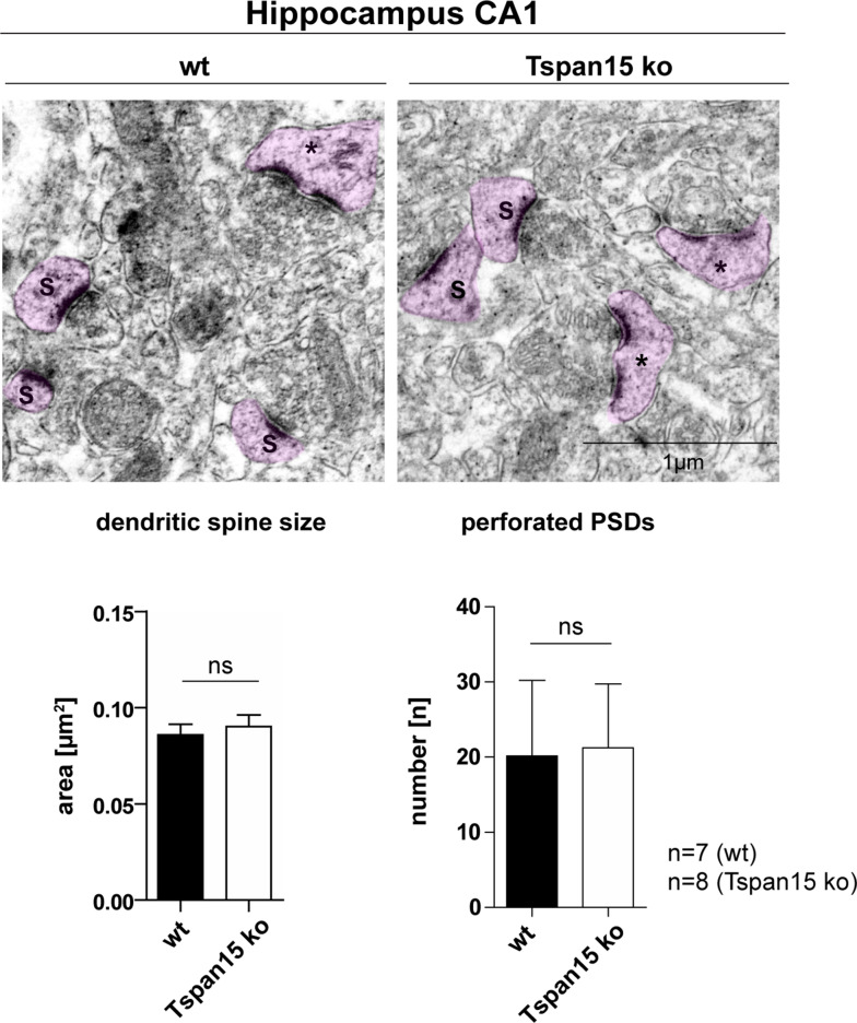
Fig. 6.
Loss of Tspan15 has no effect on the morphology of dendritic spines. Representative electron micrographs of the hippocampal CA1 region of wild-type (wt) and Tspan15-deficient mice (ko). Dendritic spines with a continuous (S) and perforated (*) postsynaptic density (PSD) are highlighted in pink. The size of dendritic spines (S) of n = 7 wild-type (wt) and n = 8 Tspan15 knockout (Tspan15 ko) animals was measured using ImageJ. Mean values ± SD of the measured spine area are shown in µm2. The number of perforated PSDs (*) of wild-type (wt) and Tspan15 knockout (ko) animals was counted. Data are shown as mean values ± SD. Statistical significance was analyzed by Student’s t test. No significant differences (ns) were observed

