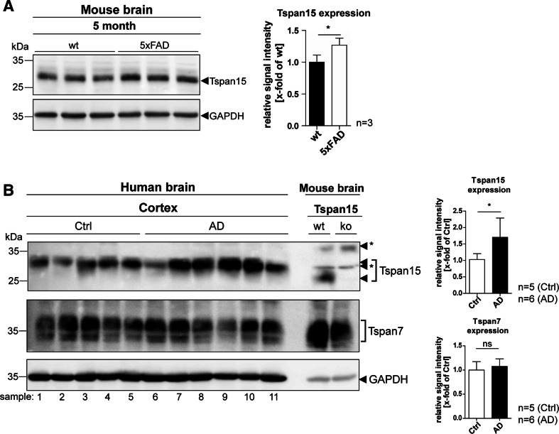
Fig. 7.
Tspan15 expression is upregulated in Alzheimer’s disease model mice and patients’ brains. a Immunoblot analysis of brain homogenates of 5-month-old wild-type (wt) and 5xFAD mice. Detection of Tspan15 (Tspan15 T2EL) and subsequent quantification of signal intensities revealed an increased Tspan15 expression in 5xFAD compared to wild-type samples. b Tspan15 expression was analyzed in post-mortem prefrontal cortex samples of human Alzheimer’s disease (AD, n = 6) patients and patients without diagnosed neurodegeneration (Ctrl, n = 5). Tspan15 wild-type (wt) and knockout (ko) brain homogenates were used to confirm specificity of the anti-Tspan15 antibody (NBP1-92540). In addition, Tspan7 expression was analyzed with a self-made anti-Tspan7 antibody. Equal protein loading was verified by GAPDH staining. Values are shown as mean ± SD. Statistical significance was tested using Student’s t test (*p < 0.05). Numbers indicate patient samples as given in Suppl. Table S1. Asterisks (*) mark unspecific signals

