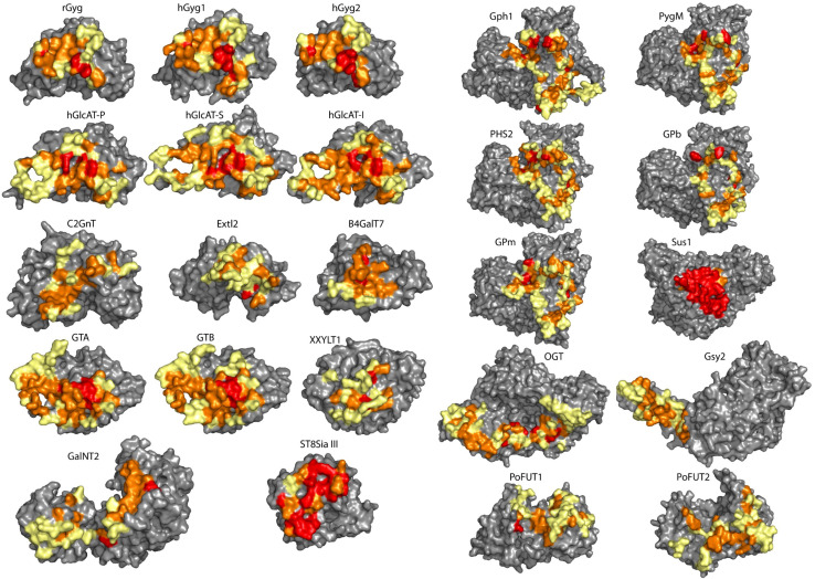
Fig. 4.
Evolutionary conservation of the amino acid sequence of the dimerization interface, visualized on each monomer (the interface facing the reader) of the 24 GTases as a colour gradient: from red (strictly conserved) through orange (high conservation) to yellow (more diverse). The residues not involved in the dimerization interface are displayed in grey. The placement of the monomers in the figure is the same as for the dimers in Fig. 1

