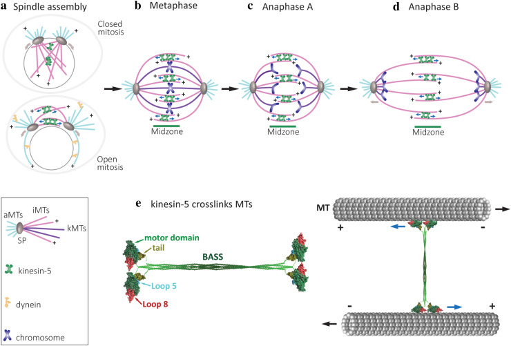
Fig. 1.
Schematic representation of the major roles of kinesin-5 motors in mitotic spindle dynamics. a Spindle-pole (SP) separation during spindle assembly in open and closed mitosis [150]. Kinesin-5 motors provide the spindle pole-separating forces by crosslinking and sliding antiparallel spindle MTs apart. The minus end-directed motor dynein (yellow shape) contributes to spindle-pole separation in cells dividing via open mitosis only [151, 152]. The direction of movement of the kinesin-5 motors and the spindle poles are indicated by the blue and brown arrows, respectively. b Metaphase, bipolar mitotic spindles with chromosomes congressed at the middle of the spindle. Three types of MTs, kinetochore MTs (kMTs, purple), astral or cytoplasmic MTs (cMTs, pink) and interpolar MTs (iMTs, light blue), are indicated by different colors. At metaphase, the chromosomes appear as pairs of sister chromatids that align in the middle of the spindle and are attached to kMTs by their kinetochores. Kinesin-5 motors crosslink antiparallel iMTs at the midzone and stabilize the spindle. In metazoans, kinesin-5 motors drive poleward flux and maintain spindle bipolarity [39, 84, 153]. c Anaphase A. Loss of cohesion between the sister chromatids marks the onset of anaphase, with sister chromatids being pulled to the opposite spindle poles. Anaphase A starts with poleward flux-based depolymerization of kMTs. d Anaphase B spindle elongation is marked by separation of the two opposing spindle poles, pulling along disjoined sister chromatids, leading to final chromosome segregation. Anaphase B results in a two- to five-fold increase in the distance between the spindle poles in both budding and fission yeast [154, 155]. Spindle elongation is mediated by cortical force generators, such as dynein motors attached to the cortex which translocate along cMTs, and kinesin-5-mediated forces produced by sliding antiparallel iMTs apart at the midzone. e Schematic presentation of a full-length kinesin-5 tetramer and its arrangement in crosslinking spindle MTs. Left: the model depicts the motor and tail domains at the end of the bipolar structure connected through the central stalk. The central stalk which mainly consists of a coiled-coil structure formed from the four helixes joining each motor domain to the corresponding tail domain, includes the bipolar-assembly (BASS) domain in the middle region, which is important for the organization of bipolar homo-tetrameric kinesin-5 complex [57, 110]. The models of Cin8 motor (aa 73–522) and tail (aa 970–1038) domains were construct by homology modelling using the Swiss Model server [152] and depicted using UCSF Chimera [155]. The motor domain is super-imposed on the cryo-electron microscopy structure of a S. pombe Cut7 motor domain-decorated MT in the AMP-PNP-bound state (PDB: 5M5I) [128]. The motor domains are represented in green, with the large loop 8 in motor domain of Cin8 (see below) in red and loop 5 in cyan; the tail domain is shown in olive green. Right: the pair of motor domains at each end interacts with the two antiparallel MTs and hence, crosslinks and slides them apart. Blue arrows indicate the direction of kinesin-5 head movement on the MTs; black arrows represent the directions of MT movement during antiparallel sliding

