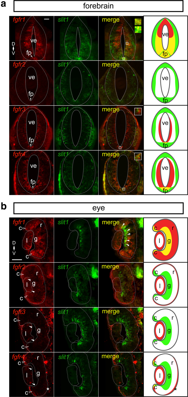
Fig. 1.
fgfr1–4 and slit1 expression patterns in Stage 32 embryos. Double fluorescent in situ hybridization on transverse sections through the forebrain (a) and the eye (b) using specific antisense riboprobes against fgfr1–4 and slit1. a There is co-expression of slit1 and fgfr1 ventrally adjacent to the ventricle and in the floor plate, fgfr3 (floor plate), and fgfr4 (floor plate). The insets focus on the regions of co-expression. b In the neural retina, slit1 co-expresses with fgfr1 in the presumptive retinal ganglion cell layer (white arrowheads) and ciliary marginal zone (unfilled arrowheads). The ciliary marginal zone (unfilled arrow heads) and the lens express different combinations of fgfrs, but not in conjunction with slit1. fgfr2–4 expression in the lens is denoted by white arrowheads. The asterisk indicates fgfr4 expression in the retinal pigmented epithelium. The dotted lines outline the boundaries of the ventricle, forebrain, lens, and neural retina. The rightmost column in each composite is a cartoon of the fgfr (red) and slit1 (green) domains with co-expression in yellow. Scale bars 50 µm. c ciliary marginal zone, fp floor plate, g retinal ganglion cell layer, l lens, r neural retina, ve ventricle

