Fig. 5.
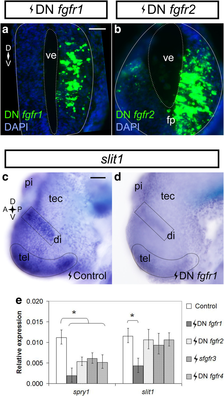
Fgfr1 inhibition downregulates slit1 in the forebrain. Stage 27/28 embryos were electroporated in the forebrain with pCS2-DNfgfr1 (a) and pCS108-DNfgfr2 (b) and processed in transverse sections for fgfr1/2 in situ hybridization at Stage 32 to reveal the expression of the constructs. In a and b, the solid outline encircles the neural tube, and the dotted outline borders the ventricle. Stage 27/28 embryos were electroporated with control pCS2-GFP (c) (n = 13 embryos from two independent experiments) or pCS2-DNfgfr1 (d) (n = 12 embryos from two independent experiments; 10/12 brains showed downregulation) and processed for slit1 expression by whole mount in situ hybridization at Stage 32. The dotted outlines in c and d indicate the slit1 domains of interest. e The slit1 expression in the brains of embryos electroporated with truncated fgfrs was measured by qPCR. spry1 was a readout of Fgfr inhibition. Bars represent mean ± SEM for n = 137 embryos from four independent experiments. *p < 0.05 statistical significance versus the control was determined using the REST 2009 algorithm. Scale bars 50 µm. di diencephalon, fp floor plate, pi pineal gland, tec optic tectum, tel telencephalon, ve ventricle

