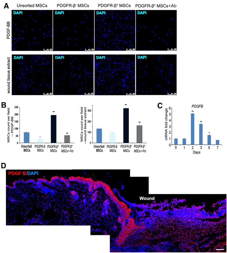
Fig. 7.
Chemotactic migration of PDGFR-β+ MSCs. a, b Transwell migration assay showed that the migration of unsorted MSCs, PDGFR-β+ MSCs and PDGFR-β− MSCs in response to PDGF-BB or fresh wound tissue extracts. Nuclei were stained with DAPI (a). The average number of cells migrated across the pores per microscopic field was quantified by Image J (b). Pre-treatment of PDGFR-β+ MSCs with a functional blocking antibody against PDGFR-β significantly reduced their migration toward PDGF-BB or the fresh wound tissue extracts (a, b). Experiments were performed in triplicate wells. c, d Real-time PCR analysis of the expression of PDGFB after wounding in mice (c). d Immunofluorescence analysis of a day-3 wound showed the expression of PDGF-B (red), which showed higher levels in the wound bed tissue and the tissue adjacent to the wound. Nuclei were stained with DAPI. Scale bars 50 µm. ***P < 0.001, **P < 0.01, *P < 0.05

