Fig. 2.
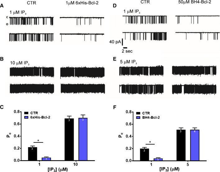
Recombinant Bcl-2 and BH4-Bcl-2 fail to inhibit IP3R activity induced by high concentrations of IP3 in single-channel measurements. a, b Representative IP3R1 single-channel recordings from DT40-3KO cells ectopically expressing IP3R1. The channel opening was evoked by 1 (a) or 10 (b) μM IP3 at 200 nM Ca2+ and 5 mM ATP, in the presence of PBS (a, b left) or in the presence of 1 μM 6xHis-Bcl-2 purified protein, which was dialysed against PBS (a, b right). c Histogram depicting the Po ± SEM for the IP3R1 under the described conditions. The total number of recordings for each condition is indicated within every bar. d, e Representative IP3R1 single-channel recordings from DT40-3KO cells ectopically expressing IP3R1. The channel opening was evoked by 1 (d) or 5 (e) μM IP3 at 200 nM Ca2+ and 5 mM ATP, in DMSO control condition (d, e left) or in presence of 50 μM BH4-Bcl-2 peptide (d, e right). f Histogram depicting the Po ± SEM for the IP3R1 under the described conditions. The total number of recordings for each condition is indicated within every bar. *Stands for p < 0.05. The single-channel recordings presented on panel d and the corresponding analyzed data on panel f for 1 μM IP3 with and without 50 μM BH4-Bcl-2 were obtained from exactly the same data set, as previously published in [39]. All other results represent newly acquired data

