Fig. 3.
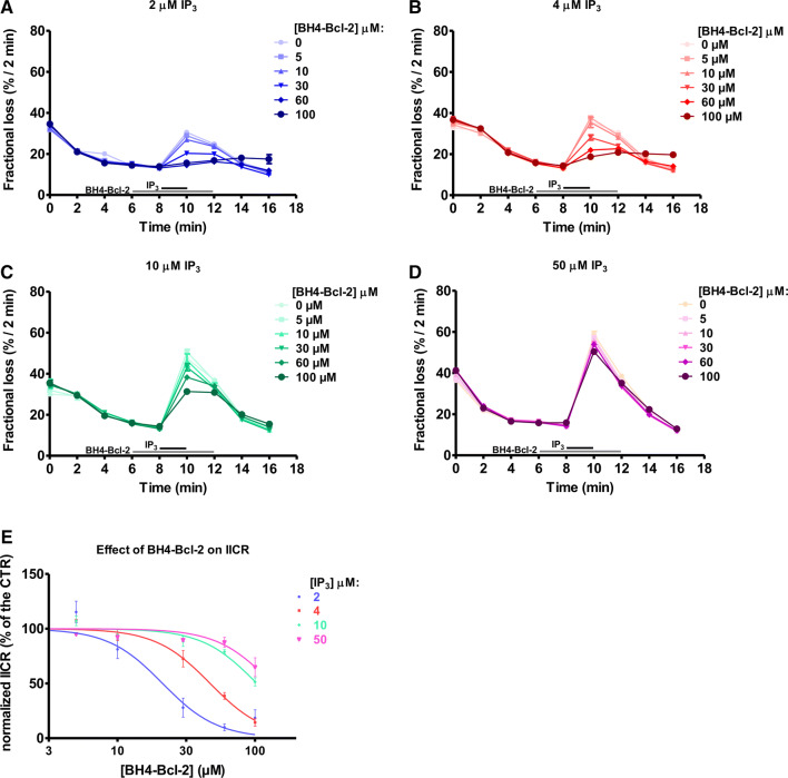
The BH4-Bcl-2 peptide fails to inhibit IICR triggered by high IP3 concentrations. a–d Typical experiment of unidirectional 45Ca2+ fluxes in permeabilized MEFs. Ca2+ release was induced by 2 (a), 4 (b), 10 (c) or 50 (d) μM IP3 (the time of addition is indicated with a black bar) in control condition or in presence of different concentrations of BH4-Bcl-2 peptide (the time of addition is indicated with a grey bar). The results are plotted as fractional loss after 2 min of incubation with IP3 minus the fractional loss before the addition of IP3 (%/2 min) as a function of time. e Concentration–response curve of the IICR as quantified from four independent experiments, performed on independently grown cell cultures. The values of IICR measured as fractional loss were calculated as percentage of the IICR in control condition (vehicle), which was set as 100%. *Stands for p < 0.05

