Fig. 1.
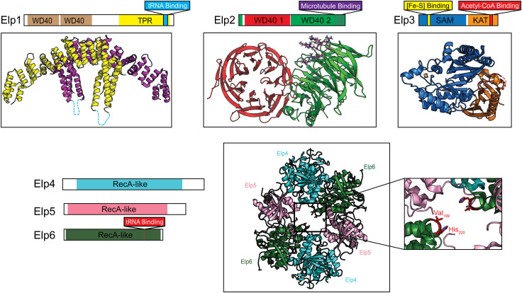
Structures of the Elp1, Elp2, and Elp3 subunits and the Elp4/5/6 subcomplex. a Schematic representation of the domain composition of Elp1 and the crystal structure of the Saccharomyces cerevisiae Elp1 dimerization domain (residues 919–1349) (PDB ID: 5CQS). Two copies of the dimerization domain are represented in yellow and purple, respectively. Basic region implicated in tRNA binding is shown in blue. b Domain structure of the Elp2 subunit and the crystal structure of full-length S. cerevisiae Elp2 protein (PDB ID: 5M2N). Residues proposed to mediate microtubule binding are shown in stick representation in purple. c Domain overview of the Elp3 subunit and the structure of Dehalococcoides mccartyi Elp3 (PDB ID: 5L7J). Acetyl-CoA binding loop and the [2Fe–2S] cluster are shown in red and yellow/orange, respectively. d Schematic representation of RecA-like domains found in Elp4 (blue), Elp5 (pink) and Elp6 (green) (left) and the crystal structure of the heterohexameric form of the subcomplex (right) (PDB ID: 4A8J). The inset shows key residues of Elp6 required for the interaction of tRNA with Elp4/5/6

