Fig. 3.
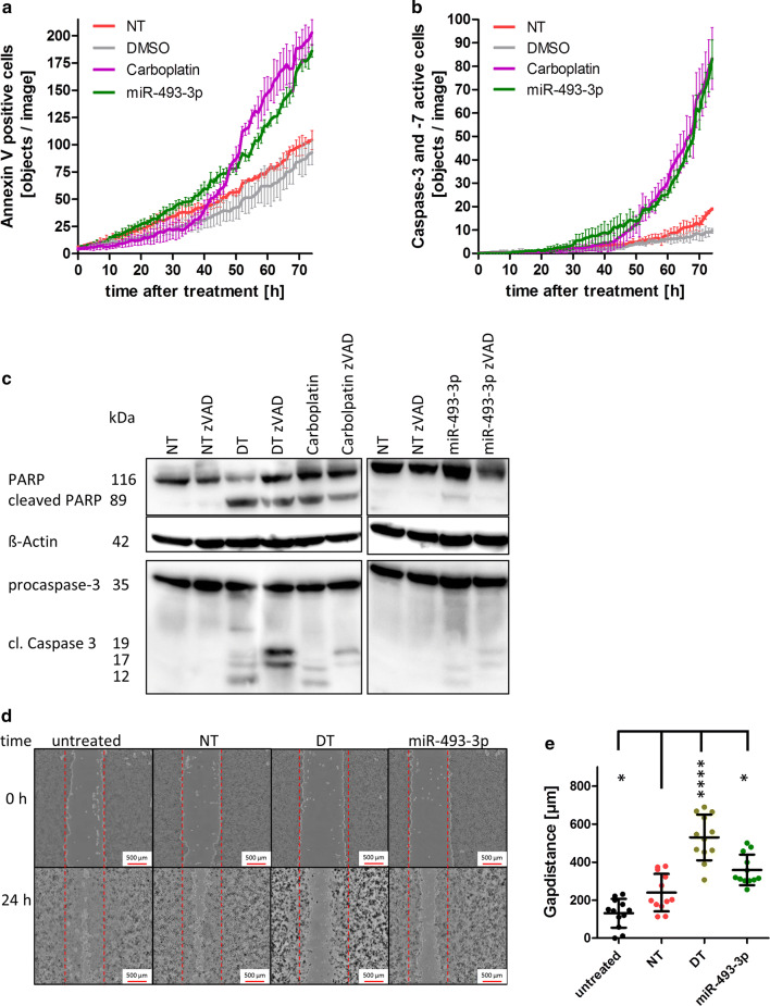
Activation of Caspase 3/-7 by miR-493. For long-term analysis, SKOV3 cells were seeded and transfected as described in Fig. 2. To detect apoptosis induction by miR-493, the cells were stained with IncuCyte AnnexinV Red Reagent (a) or IncuCyte Caspase-3/7 Green Apoptosis Assay Reagent (b). The cells were automatically photographed every hour by the IncuCyte ZOOM System. The amount of AnnexinV-positive cells or cells with activated Caspase 3 or -7 was calculated by the IncuCyte ZOOM Software. For Western Blot analysis, the cells were harvested 72 h after treatment. ß-Actin served as a loading control. Cells were treated with 30 µM zVAD to inhibit Caspase 3 activity (c). For measuring motility, cells were seeded, scratched with a 1000 µl pipet tip and transfected with the negative control siRNA (NT), miR-493-3p or the cell death positive control (DT). 0 h and 24 h after transfection images of the cell layer were taken by the automated single well microscope NyOne (d). The distance between the gap was measured and plotted on a box-and-whisker diagram (e). Statistical analyses were performed by one-way ANOVA followed by Bonferroni post-test. [n = 3 replicates; mean ± SD, *p < 0.05, **p < 0.01, ***p < 0.001, ****p < 0.0001]

