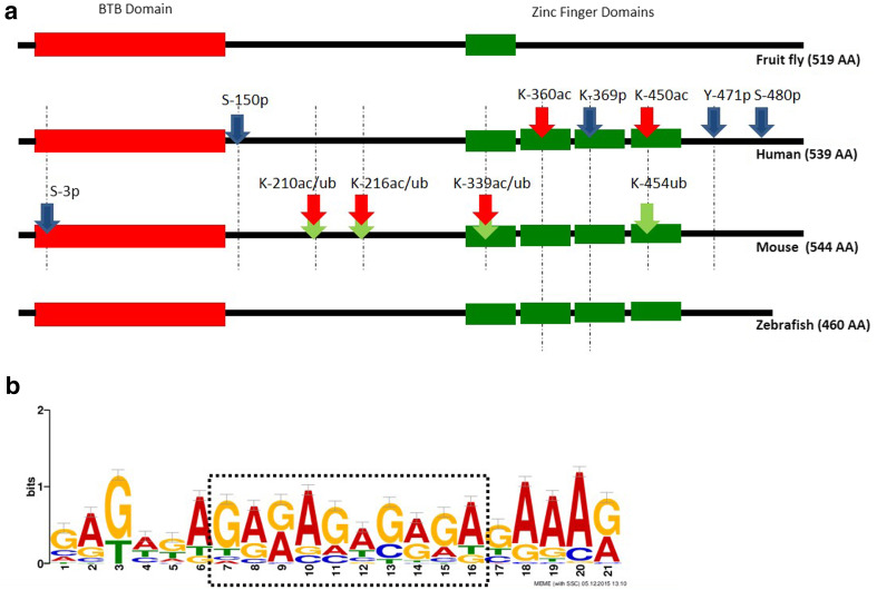
Fig. 1.
a GAF domain structure and post-translational modifications: all GAF proteins contain a BTB domain (red); DmGAF contains one zinc finger domain, while vGAF contains four zinc finger domains (green). PTM site phosphorylation (blue), acetylation (red), and ubiquitination (green) are shown by arrows marked with amino acid residue position. Dotted lines show the extent of conservation of these amino acid residue in human, mouse and zebrafish proteins. b The DNA-binding motif of vGAF proteins. Thirty-three known binding sites of vGAF (ThPOK) in mouse genome were taken from literature [21, 39, 51]. MEME analysis done on these sequences shows a 21 bp long binding motif with a central polypurine-rich sequence

