Fig. 1.
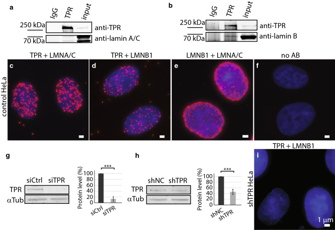
TPR co-immunoprecipitates with lamin B1. a Immunoprecipitation of lamin A/C by anti-TPR antibody. b Immunoprecipitation of lamin B1 by anti-TPR antibody. Proximity ligation assay shows the close distance between TPR and lamin A/C (c), TPR and lamin B1 (d), lamin B1 and lamin A/C (e) and negative control with no primary antibody used (f). Western blotting and quantification show the reduction of TPR amount in HeLa cells with siTPR for 48 h: Student t test, T = 12.35, df = 5, P < 0.001 (g) or in stable HeLa cell line expressing shTPR: Student t test, T = 11.01, df = 7, P < 0.001. Graphs represent data from three independent biological replicates. 3.5 × 10⁶ cells were used as a starting material. 10 μg of total protein per each lane was loaded for the analysis (h). i Proximity ligation assay detected reduced abundance of the TPR–lamin B1-positive signal in Hela cells with reduced TPR protein levels by shRNA. Scale bar 1 µm. ***P < 0.001

