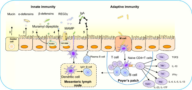
Fig. 3.
Immune mechanisms for limiting the commensals within the epithelial layer. Innate immunity: (1) mucin glycoproteins of the goblet cells form a layer over the epithelia to restrict bacterial adhesion; (2) constitutive expression of α-defensins from epithelial cells without microbial signal; (3_ β-defensin expression by muramyl dipeptide of Gram positive bacteria mediated by cytosolic nucleotide-binding oligomerization domain-containing protein 2 (NOD2); (4) C-type lectin regenerating islet-derived protein 3γ (REG3γ) via microorganism-associated molecular patterns (MAMP) activates toll-like receptors (TLRs) recruiting cytosolic adaptor myeloid differentiation primary-response protein 88 (MYD88) to restrict bacterial penetration through epithelial layer; (5) dendritic cells (DC) located beneath the epithelial dome of Peyer’s patches take up bacteria and migrate to mesenteric lymph node and induce B cells to differentiate into IgA+ plasma cells for secretion of IgA. Transcytosis of IgA across the epithelial cell layer limits bacterial association with the epithelium. Adaptive immunity: transcytosis of bacteria through M cells and luminal antigen activates DC in the Peyer′s patch and helps to differentiate the naive CD4+ T cells into Treg and TR1 characterized by expression of anti-inflammatory cytokines transforming growth factor (TGFβ) and IL10. The TH1, TH2, and TH17 cells are characterized by proinflammatory cytokines including (1) IFNγ, (2) IL-4, 5 and 13 and (3) IL-23 and IL-17F, respectively

