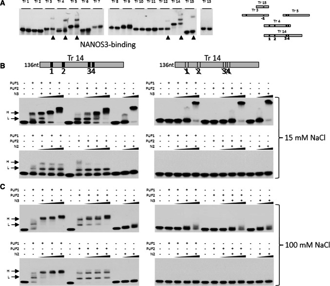
Fig. 4.
NANOS2 and NANOS3 bind to SIAH1 3′UTR and form complexes with PUF domains. a EMSA analysis of all the SIAH1 transcripts for testing binding with NANOS3. Numbers correspond to SIAH1 transcripts; the left lane represents migration of the naked RNA of each fragment, whereas the right lane represents its migration in the presence of NANOS3 protein. Lanes showing retardation bands are indicated with an arrow. b EMSA analysis of SIAH1 3′UTR transcript 14 (Tr 14, wild-type or mutated) for binding PUF1 or PUF2 domain alone or in combination with NANOS3 or NANOS2 in 15 mM NaCl. NANOS proteins were used at a concentration of 5–15 μM, whereas PUF1 and PUF2 were used at 100 nM. Wild-type Tr 14 is on the left and mutated Tr 14 is on the right. c EMSA analysis according to the same scheme as b, but performed at a higher NaCl concentration (100 mM). H higher complex; L lower complex

