Fig. 3.
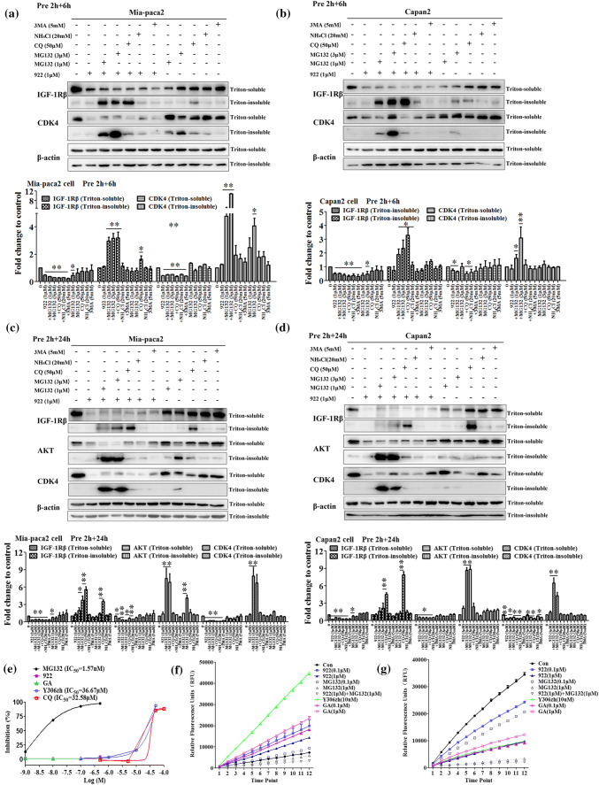
NVP-AUY922 induces IGF-1Rβ degradation mainly via the lysosomal pathway. a–d Mia-paca2 and Capan-2 cells were pretreated with MG132 (1, 3 μM), CQ (50 μM), NH4Cl (20 mM) or 3 MA (5 mM) for 2 h and then treated with 1 μM of 922 for an additional 6 h or 24 h. The IGF-1Rβ, CDK4 or AKT protein levels from Triton-soluble and Triton-insoluble fractions were measured by western blotting. β-actin was used as a loading control. Relative protein levels were quantified by densitometry and are shown in the histogram. e The chymotrypsin-like proteasome activity was determined as a magnitude of fluorogenic proteasome substrate (Suc-LLVY-AMC) degradation. Human 20S proteasome from Mia-paca2 cell lysates was incubated with 922, GA, Y306zh, CQ, or MG132, and the chymotrypsin-like proteasome activities were monitored after 60 min. MG132 served as a positive control. Relative proteasome activity is represented by the percentage of fluorescence compared with the control. f, g Mia-paca2 cells were treated with the indicated doses of 922, GA, Y306zh, CQ, or MG132 for 6 h or 24 h, and the chymotrypsin-like proteasome activities were monitored every 10 min. All data are expressed as the mean ± SD (n = 3). *P ≤ 0.05, **P ≤ 0.01 versus con

